
Magnified Dentistry
Loupes magnification or microscopes are used with all dental work. Dr. Mamoun believes that dental work cannot be optimally performed without magnification. In addition, supplementary head-mounted lighting provides superior lighting compared to the standard overhead dental chair light.
Dr. Mamoun focuses on using microscope-level magnification to help ensure extreme precision and control while performing dental work, and has written numerous articles in dental journals about applications of microscopes when performing dental procedures.
Most dental work happens at a very small scale: the edge where a filling or crown meets the tooth, a hairline crack, the opening of a root canal. Magnification enlarges that working field, and light aimed along the dentist’s line of sight lets the enlarged view be seen clearly.
For you in the chair, little changes: loupes and a headlight are worn by the dentist, and a microscope sits above the chair on a movable arm.
Loupes
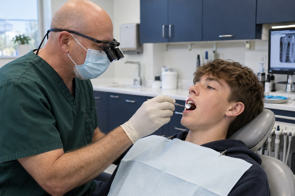
Dental loupes are small binocular telescopes built into a pair of glasses and focused at the distance the dentist works from the patient’s mouth. They enlarge the view several times over while leaving both hands free, and they move wherever the dentist moves.
Dr. Mamoun set out the case for them in his 2009 article in General Dentistry, “A rationale for the use of high-powered magnification or microscopes in general dentistry.”
Operating microscopes
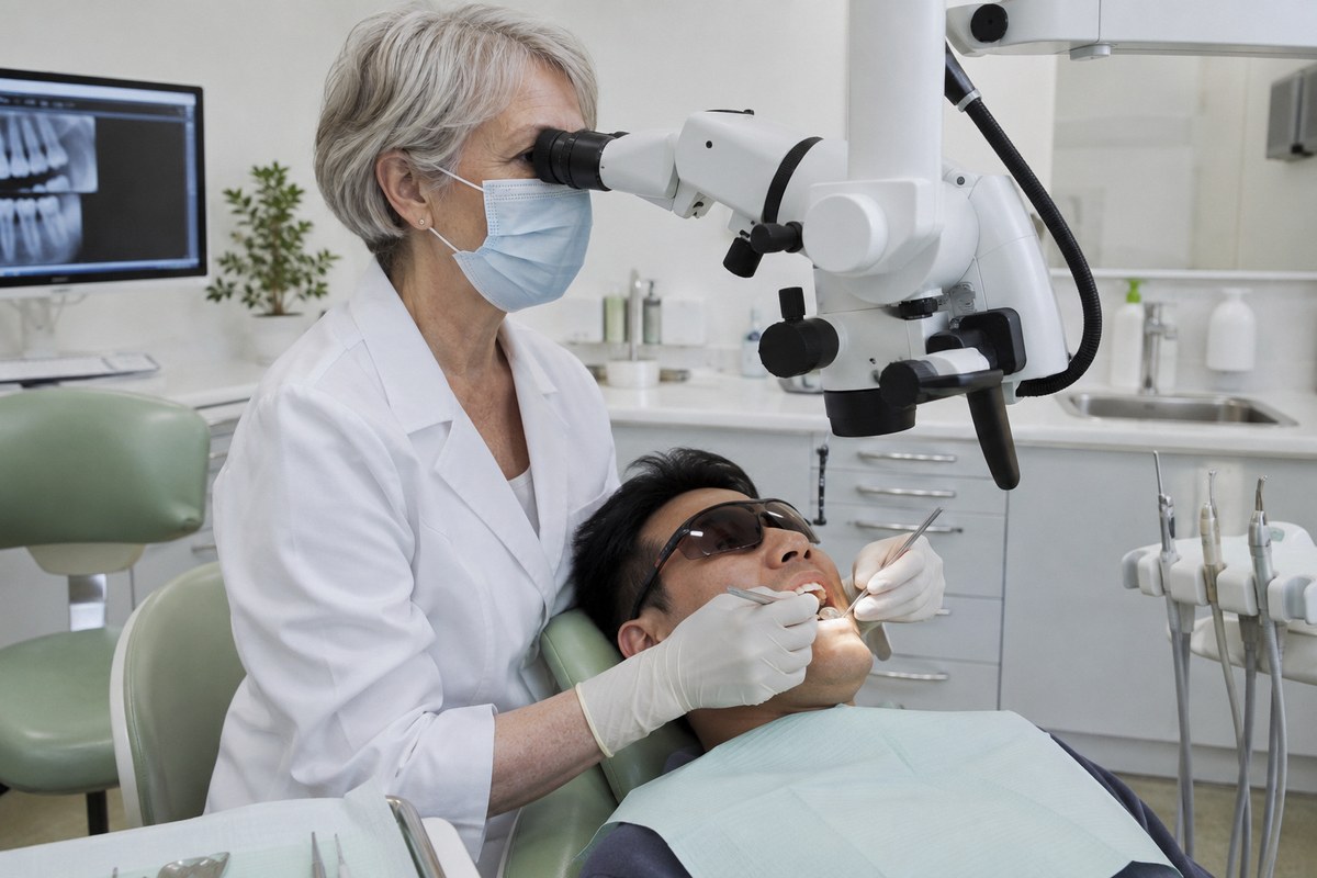
A dental operating microscope stands beside the chair on an articulated arm and swings over the patient. It magnifies further than loupes, over an adjustable range, and carries its own light through the optics, so the light falls exactly where the dentist is looking.
Dr. Mamoun has published microscope-based approaches for fitting crowns and bridges, for designing removable partial dentures and for opening the root canals of upper molars.
Read his microscope-based approach to root canal access on PubMed
Head-mounted lighting
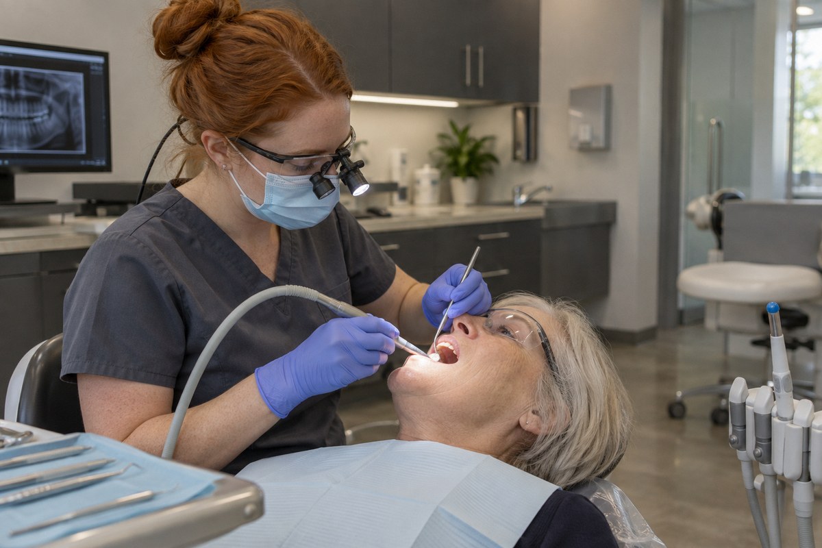
A head-mounted light is a small, bright lamp worn on the loupes or on a headband. Because it points wherever the dentist looks, its beam stays in line with the view, and the dentist’s own head and hands do not shadow the tooth the way they can under an overhead light.
His published research
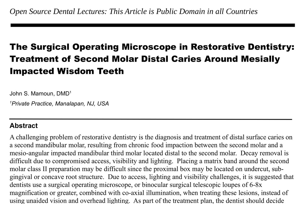
In April 2013 Dr. Mamoun published a public-domain lecture, “The Surgical Operating Microscope in Restorative Dentistry: Treatment of Second Molar Distal Caries Around Mesially Impacted Wisdom Teeth.” It recommends a surgical operating microscope, or loupes of 6 to 8 times magnification or greater with co-axial illumination, for decay that is hard to reach, see and light.
He has written about loupes and microscopes across his publishing career. Among his articles indexed on PubMed:
- A rationale for the use of high-powered magnification or microscopes in general dentistry. General Dentistry, 2009. PubMed
- Use of high-magnification loupes or surgical operating microscope when performing dental extractions. New York State Dental Journal, 2013. PubMed
- Use of high-magnification loupes or surgical operating microscope when performing prophylaxes, scaling or root planing procedures. New York State Dental Journal, 2013. PubMed
- Using microscopes in fixed prosthodontics: try-in, adjustment, and insertion of crowns and bridges. Dentistry Today, 2014. PubMed
- The path of placement of a removable partial denture: a microscope based approach to survey and design. The Journal of Advanced Prosthodontics, 2015. PubMed
- Preparing and restoring composite resin restorations: the advantage of high magnification loupes or the dental surgical operating microscope. New York State Dental Journal, 2015. PubMed
- The maxillary molar endodontic access opening: a microscope-based approach. European Journal of Dentistry, 2016. PubMed
Loupes or a microscope are used with all dental work at Sunbeam Dental. If you have a question about how magnification applies to your own treatment, call us at (732) 431-2888.